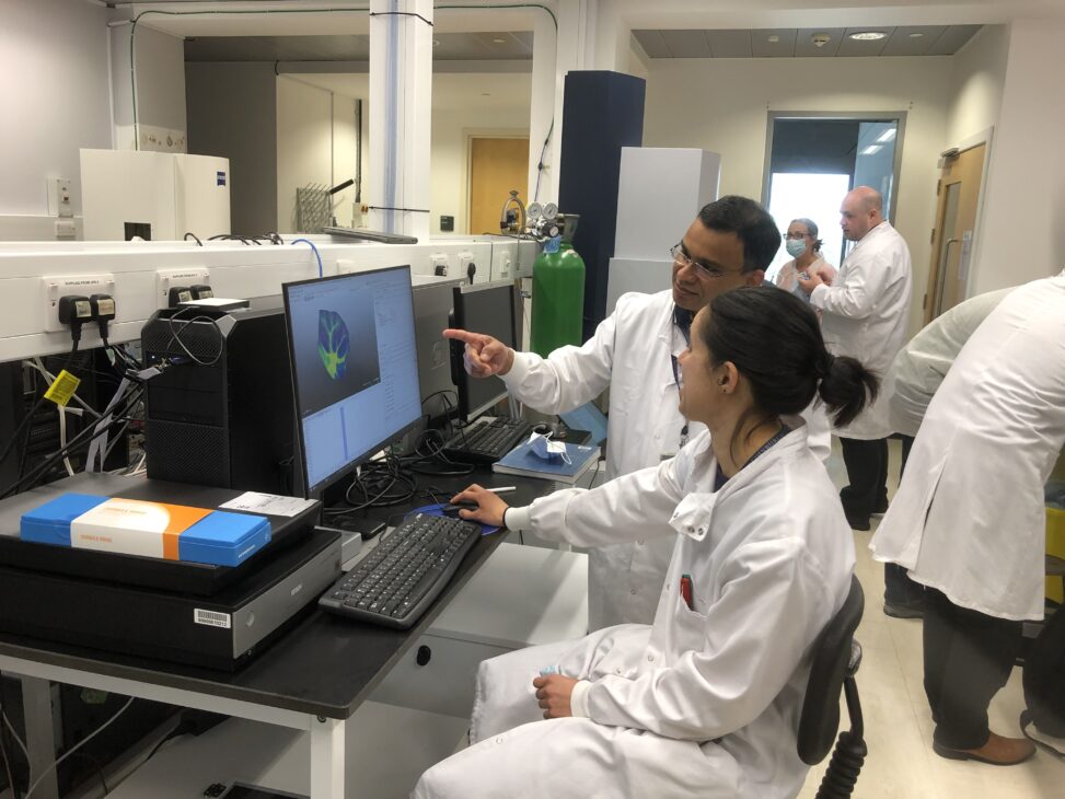
New laboratory facilities at the Roslin Institute are allowing fast, precise analysis to identify protein molecules of interest in cells, tissues or body fluid.
Our understanding of disease benefits enormously from insight into proteins. As the building blocks of tissues, organs and systems, these large, complex molecules are at the heart of biological organisms and are central to the functional processes that enable and sustain life.
Understanding their physical make-up and structure, abundance, and how they interact with other molecules, is important for our knowledge of what constitutes healthy tissue, and the impacts of disease.
In a living organism, the types of proteins present can vary from cell to cell and at various times, presenting a challenge in characterising and understanding their functions.
The newly- installed laboratory facilities at the Roslin Institute are allowing fast, precise analysis to identify protein molecules of interest in cells, tissues or body fluid. The Proteomics and Metabolomics Facility can support a broad range of studies of proteins in various tissues by offering detailed analysis of the proteins present, and is available to scientists across the University.
Protein signatures
The facility’s mass spectrometer, one of only two of its type in the UK, works via a laser light that interacts with proteins in samples. It generates images detailing the molecular fingerprint for a range of proteins, and their locations in situ at a high spatial resolution– up to 10 micrometers, or one thousandth of a cm – the equivalent of a cell nucleus.
Where in-depth understanding of sample composition is required, biological samples may be prepared and delivered to the mass spectrometer through a device that can separate the components of interest before their mass is analysed in the mass spectrometer.
The instrumentation can process up to 300 samples in one day, allowing researchers to quickly generate a wealth of data on various proteins of interest.
Mass spectrometry presents global picture of all detectable proteins present in a sample. This contrasts with conventional analysis using microscopy imaging, in which the proteins to be analysed must be decided ahead of sample preparation. Mass spectrometry imaging can also pinpoint small molecular components of the cells such as products from metabolic processes, lipids or even administered drugs. Where individual components have identical mass, they can be distinguished by shape.
“This means studies can be discovery-led rather than hypothesis-led – we don’t have to decide in advance what we expect to find in damaged tissue,” explains Dr Tom Wishart, Proteomics and Metabolomics Facility Academic Lead. “For example, in a brain affected by Alzheimer’s disease, we can compare tissue from areas of the brain affected by disease against healthy tissue, to identify the nature of the proteins involved, as well as just seeing where the damage physically occurs.”
The highly sensitive instrumentations are suitable for projects where only small quantities of precious tissue are available, such as biopsy or post-mortem studies.
Real time analysis
Also available in the proteomics lab is Direct Analysis in Real Time (DART) mass spectroscopy. This device, which is unique in an academic laboratory in Scotland, works with solid or liquid samples without the need for any specific preparation.
It has many potential applications in forensics, and has been applied to a study seeking to distinguish between bone tissue from wild or captive-bred big cats, in a study that supports efforts to tackle poaching.
In addition to these capabilities, researchers are bringing into operation an electron microscope that can work with 3D samples. The device repeatedly slices and scans tissue blocks from samples, to create a three-dimensional image in ultra-high resolution. The only device of its type in Scotland, this serial section blockface scanning electron microscope can be applied, for example, to examine cell membranes, viral particles, or looking at connections in the brain.
The Proteomics and Metabolomics facility in Roslin represents a high-level service beyond the reach of many individual labs, explains Dr Dominic Kurian, Proteomics and Metabolomics Facility Manager.
“We can support biomedical or clinical research by offering capabilities that exceed those available within individual groups. We’re actively seeking to develop new ways of applying the technologies to support research and expand the scope of the facility, and we’d be glad to hear from colleagues whose studies might benefit from novel use of the technologies.”
Catriona Kelly, science writer and editor, Easter Bush campus.


Leave a Reply