What is a lichen?
This is the audio for this halt. The text below is a transcription of the audio! The audios do not contain all the content written in the text to keep the audios engaging. If you want to have more details, check out the text. Enjoy!
“In the Western tradition there is a recognized hierarchy of beings, with, of course, the human being on top—the pinnacle of evolution, the darling of Creation—and the plants at the bottom. But in Native ways of knowing, human people are often referred to as “the younger brothers of Creation.” We say that humans have the least experience with how to live and thus the most to learn—we must look to our teachers among the other species for guidance. Their wisdom is apparent in the way that they live. They teach us by example. They’ve been on the earth far longer than we have been and have had time to figure things out.”
Robin Wall Kimmerer in Braiding Sweetgrass: Indigenous Wisdom, Scientific Knowledge, and the Teachings of Plants
I would like to take you through a world of questions, entanglements, and connections. A world of mystery and imagination. A world where little is known. Through this adventure and journey of lichen discovery, I would like to imagine with you other stories about us, our perceptions, our beliefs and our futures… about the cities. I would like to open a door to another world, a world where we create spaces – like cities – by considering other organisms, like lichens. I would like to defy the division between nature/culture or between human/more-than-human often found in environmentalist movements by highlighting the intricately intertwined relationship between living beings through co-development/co-evolution processes. I hope that through this discovery you will feel the interconnections between all beings, even in an urban environment.
Lichens (pronounced L-I-K-E-N), as we will see, push us to these reflections and point out to the fact that our bodies do not end at our skin. Lichens invite us to consider new behaviours and new sensitivities. By observing one of the multiplicities by which living organisms can experience the world around us I hope you will travel into other imaginaries.
I suggest you follow me for a few hours of discovery of an organism and another way of living and feeling the world. For this, I ask you to listen, to perceive, to open your senses.
Before you start, I could like you to close your eyes. Feel the ground below your feet, sense the breeze or the strong wind (as it often happens in Edinburgh) on your skin. Can you hear the soundscape of the place where you are standing? Is it busy? Can you hear the variety of living beings around? Some humans chatting, some birds singing, some leaves moving by the wind.
Now imagine mushroom hyphae. 🍄 If you do not know what hyphae are, let me introduce you to these incredible living structures.
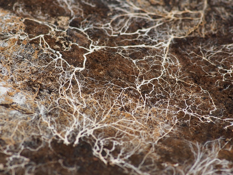
Hyphae are long, white, earthy filaments and form the structure of fungi. Assembled together in a decentralised and anarchic fashion, hyphae form the mycelium. The mycelium is not a thing, it is a process. It is constantly being created and in movement. Water and nutrients flow through the networks of mycelia which can be stimulated electrically. Some fungi therefore conduct electricity along their hyphae, similar to the impulses of nerve cells in animals.
Despite their delicacy, hyphae have incredible strength; they push the earth, but also sometimes cement (like Coprinus comatus). Imagine these hyphae in the earth, under your feet, in this bubbling ecosystem, surrounding the roots of plants. Hyphae (of the mycorrhizae fungi type) often surround the plant root and create an exchange, bringing nutrients to the trees or plants and taking carbohydrates produced from photosynthesis in exchange. I suggest you watch this video by Merlin Sheldrake which is a laser analysis of the relationship between the hyphae of a fungus and a plant. The hyphae also form the structure of the mushroom we eat. And, according to some estimates, if you take a piece of mycelium found in a teaspoon of soil and stretch it out, it could be anywhere from a few hundred metres to kilometres long.
Now imagine an exchange between the hyphae of fungi that we talked about and algae. Not the algae you might see on the beach, but small cells.
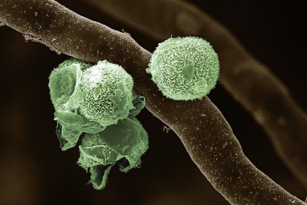
In the lichen, the hyphae are not alone, they are entangled in and with alga cells or cyanobacteria. These photosynthetic structures – algae and cyanobacteria – use the energy of the sun as well as carbon dioxide (CO2) to produce their energy. This cooperation between photosynthetic and fungal forms creates the lichen. This intimate relationship of cooperation (called a symbiosis) allows both organisms to live in places where they could not have lived alone.
Lichens are worlds, they are ecosystems. They are inhabited by hundreds if not thousands of other species including fungi and a myriad of bacteria (Pingle, 2017).
So how do we define these organisms that form an assemblage of many species, that are not “one” but are at the same time. Where is the boundary of the lichen? What is a lichen?
A lichen is a symbiotic organism. This means that it is an intimate relationship between different species where one organism is considered the host and the other is the symbiont. Symbiosis is a spectrum that contains several kinds of relationships between living organisms, including mutualism on the one hand and parasitism on the other.
Mutualism is a relationship where both organisms benefit from the association. For example, humans depend on their microbiome – which is the community of microbes in the gut and their genetic material – to digest food. Some research also shows that the microbiome in our gut can affect our immune system and moods. On the other hand, the microbes, through the symbiosis, live in an environment where they have access to food and are protected. Both organisms (human and microbe) benefit from this relationship.
In a parasitic relationship, one organism benefits from the relationship while the other organism loses. The SARS-CoV-2 virus is a parasite for humans and generates a disease that we all know by now: the coronavirus. The virus, on the other hand, finds a comfortable environment in humans where it can reproduce.
Well, back to our lichens….
A lichen is a stable and lasting mutualistic association between fungi and an alga or cyanobacterium.
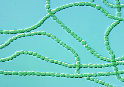
Cyanobacteria look like algae but are actually single-celled or colonial bacteria (see photo).
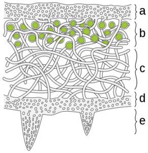
The diagram above represents a cross section of the thallus. The algal cells are entangled in the fungal hyphae (part b). The thallus is the visible surface of the lichen and consists of several layers of cells. The thallus is a word I suggest you remember, as it is an essential characteristic for identifying the lichen.
The upper and lower parts of the diagram represent the cortices (a, upper cortex and d, lower cortex) that protect lichens from their environment. These structures are primarily formed of fungal (mushroom) tissues. At the bottom of the upper cortex is the layer of algae or cyanobacteria when they are in symbiosis with the fungi (represented by the green circles in part b). Then comes the medulla, it is the layer of mushroom hyphae (part c on the diagram). The part e represents the rhizines, which lichens use to fix themselves to the surface on which they are found.
With the naked eye, we can only see the thallus as well as the lower and upper cortex. If you have access to a microscope, I suggest you take a piece of thallus and cut it in half. You will then be able to see the green layer, the algae, as well as the white medulla.
Now that we know the anatomy of the lichen, let’s move on to their functions!
Algae (and cyanobacteria) are photosynthetic organisms. The algae and/or the cyanobacteria are called photobionts. The photobionts capture the energy from the sun, and with water and carbon dioxide (CO2) from the air, they transform it into carbohydrates. Carbohydrates are sugars that provide energy to the lichens that are partly absorbed by the fungal cells.
The fungus, on the other hand, provides a protected environment and gathers moisture, which prevents the algal cells from drying out. The fungus also provides the algae or cyanobacteria with nutrients. Lichens are always named after the dominant fungus. This will become important when we start learning the names of the different lichens.
Algae and cyanobacteria can live without being associated with fungi, but their symbiotic association with the fungus changes them radically (structurally and morphologically).
Note 1: Photosynthetic organisms are autotrophic – they produce their own energy source. We humans get our energy from the food we eat, because we cannot photosynthesise – we are heterotrophs.
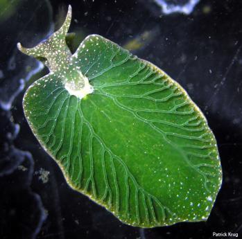
Note 2: Normally, we think of plants as photosynthetic organisms. However, some animals produce their energy from the sun’s energy. Have you ever heard of Elysia chlorotica? This sea slug is found on the east coast of the United States and has the ability to sequester a chloroplast – the organelle where the process of photosynthesis takes place – from its prey, an alga. This non-permanent symbiosis allows the sea slug to survive for up to 12 months thanks to the energy supply that the algae provides through photosynthesis.
Activity 🌸
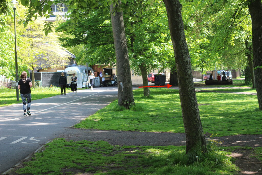
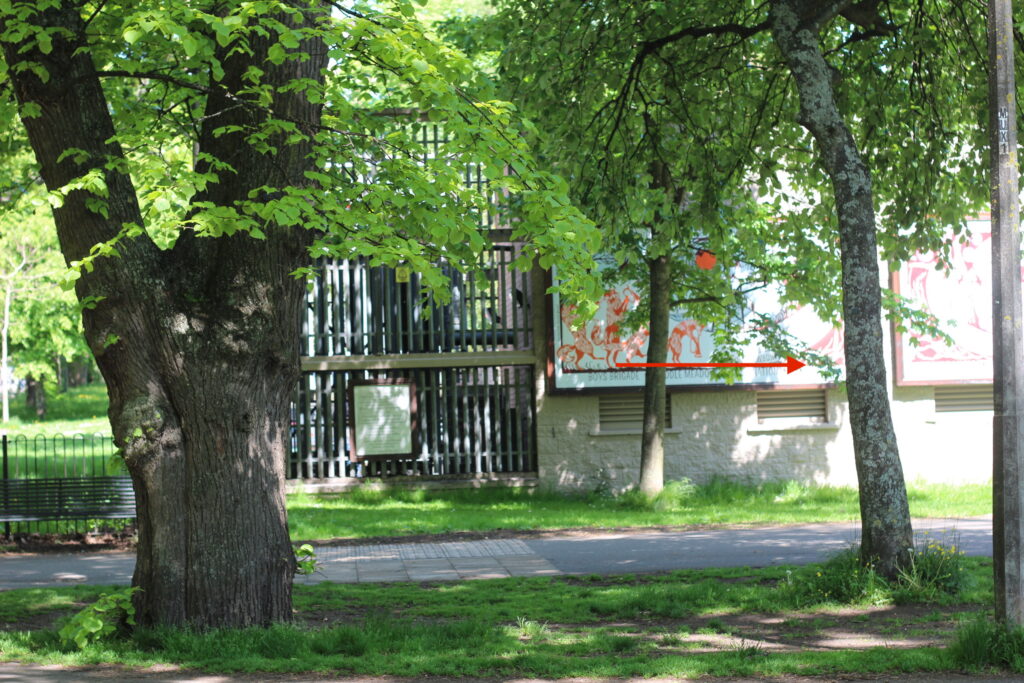
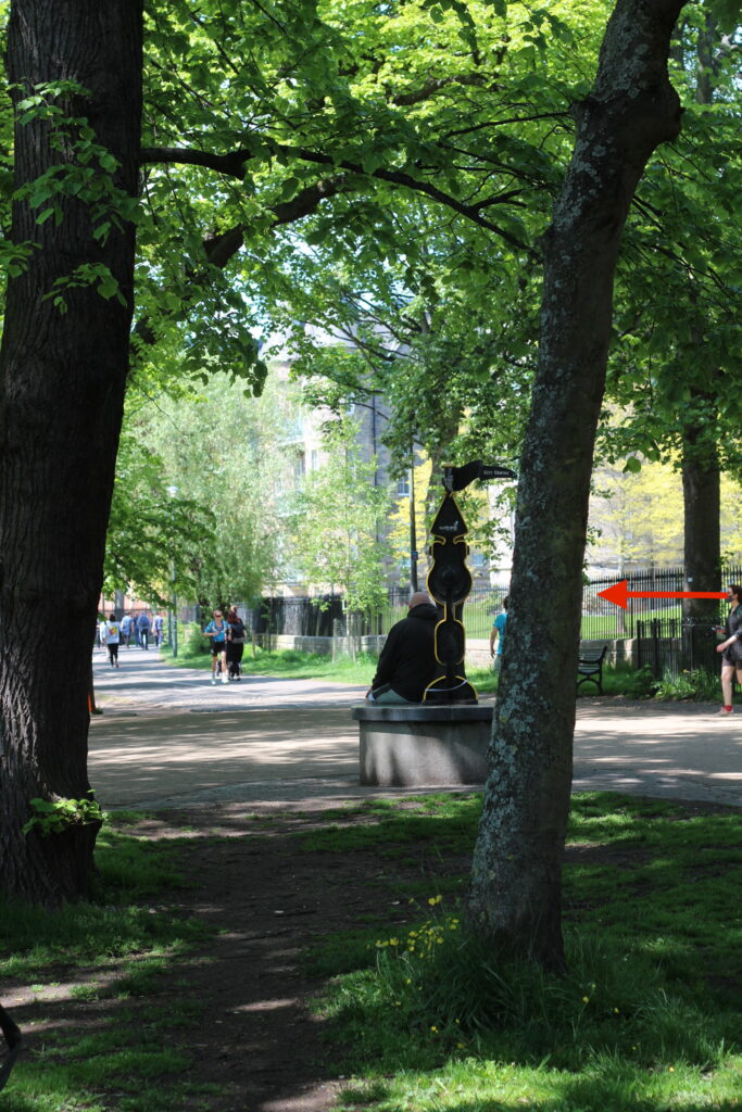
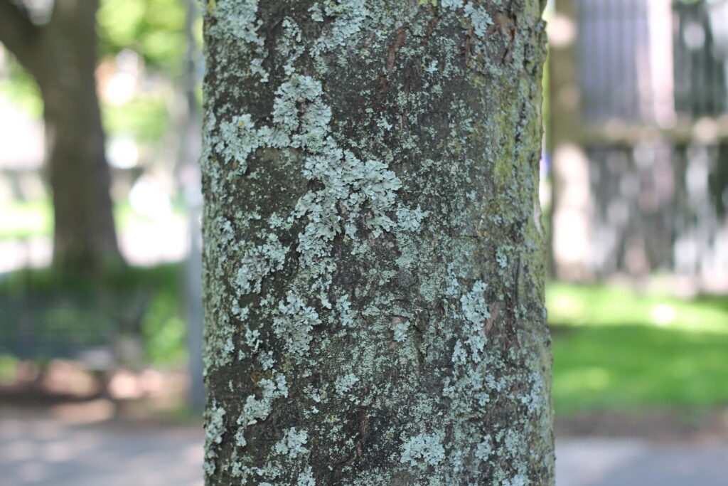
Have you found the tree we will be looking at to start with?
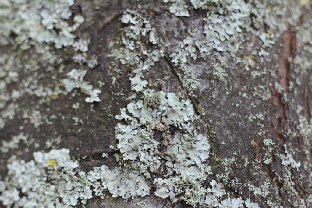
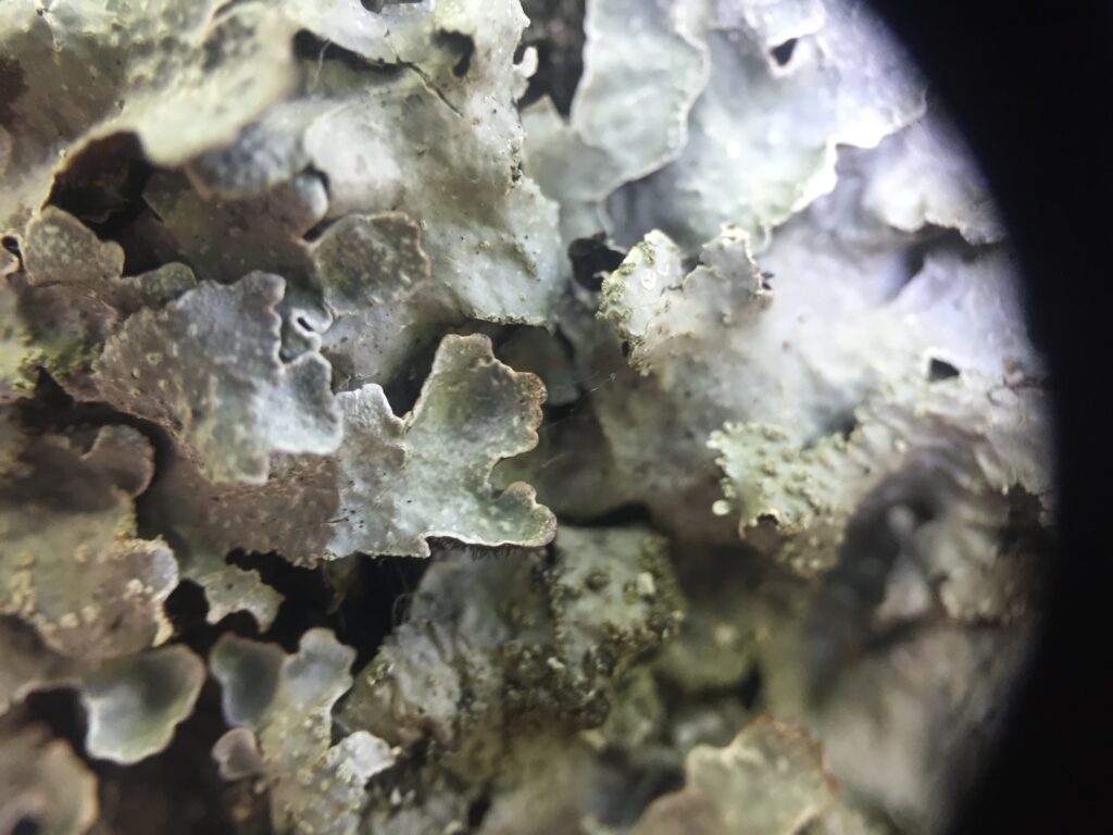
On the tree in front of you, can you see the lichens?
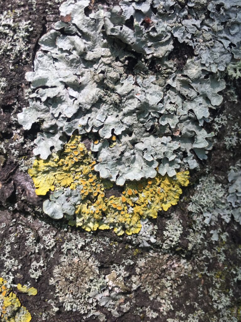
Look at the pictures below to help you.
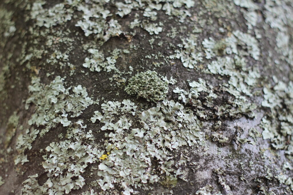
The forms of lichens are very varied.
Can you already differentiate between different morphological forms ? Different colours ?
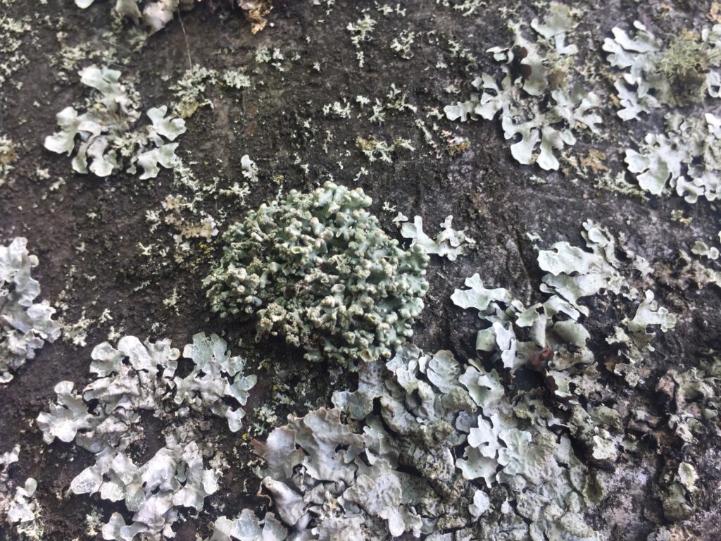
We will learn a few of those as we go but below, you can find some pictures of different species.
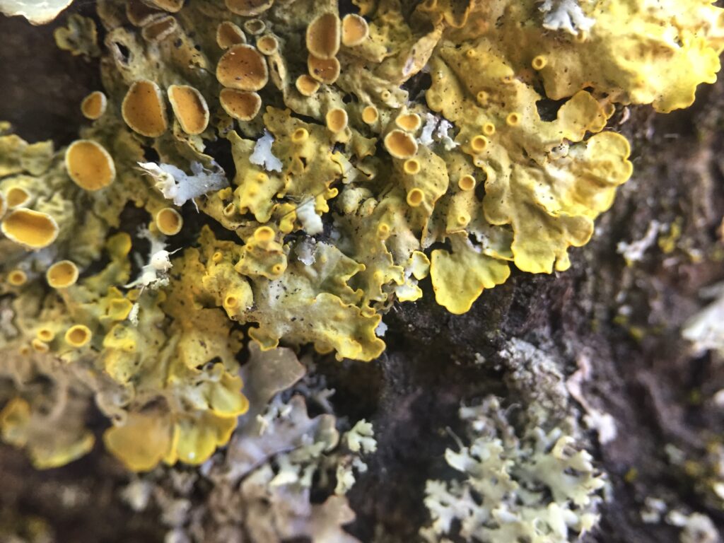
Here are some pictures of the different individuals you might see on the tree in front of you.

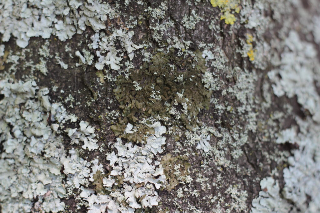
A common lichen (the bright yellow one), Xanthoria parietina, that we will see a lot and talk more about later on this walk.
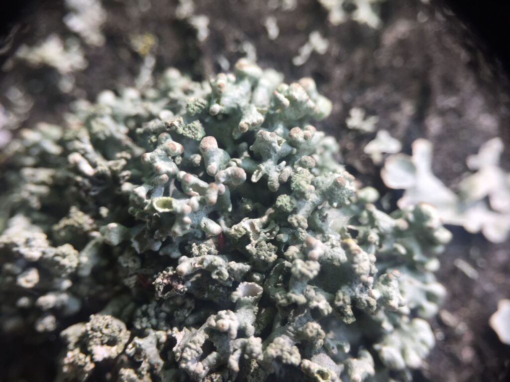
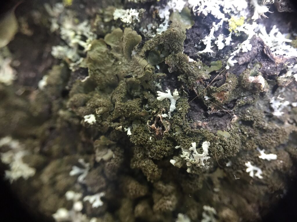
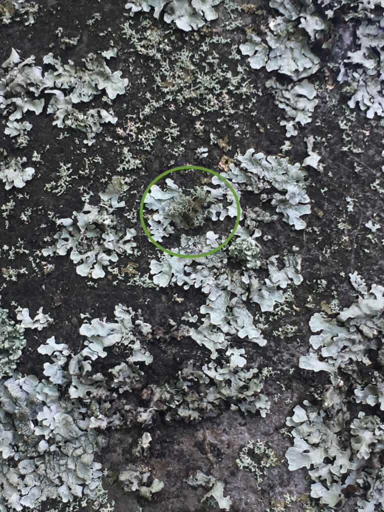
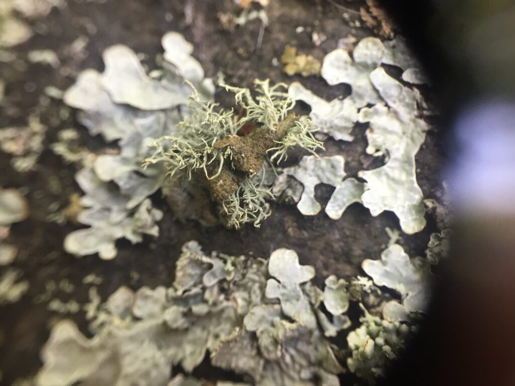
Lastly, I would like to share a special discovery with you. When I was looking at the lichens on this tree, I spotted a lichen of the genus Usnea. This genus is normally present in less polluted environments. It was a surprise to find this individual in the middle of the city. The Usnea spp. can be recognised by their long filaments (lobes) that can be stretched out a lot before breaking. These are fruticose lichens. We will talk about what it is at the next halt.
If you are already more experienced, you can look at the identification key available here and try to identify the different species.
References
- The content of this walk is very much inspired by the book of Merlin Sheldrake:
Sheldrake, M. (2020). Entangled life: how fungi make our worlds, change our minds & shape our futures. Random House.
You can also find amazing videos about fungi on his YouTube account here.
- Pringle, A. (2017). Establishing new worlds: the lichens of Petersham. Arts of Living on a Damaged Planet.
Feedback
If you don’t continue the ride, can you give us feedback on your experience here? It will help us improve!
That’s the end of this first halt, we meet at the next halt – at the Pavilion on the Meadows park at the pedestrian crossing on Melville Drive. The exact location can be found on the map here! 👇🏾👇🏾