Previous Webinars
2021
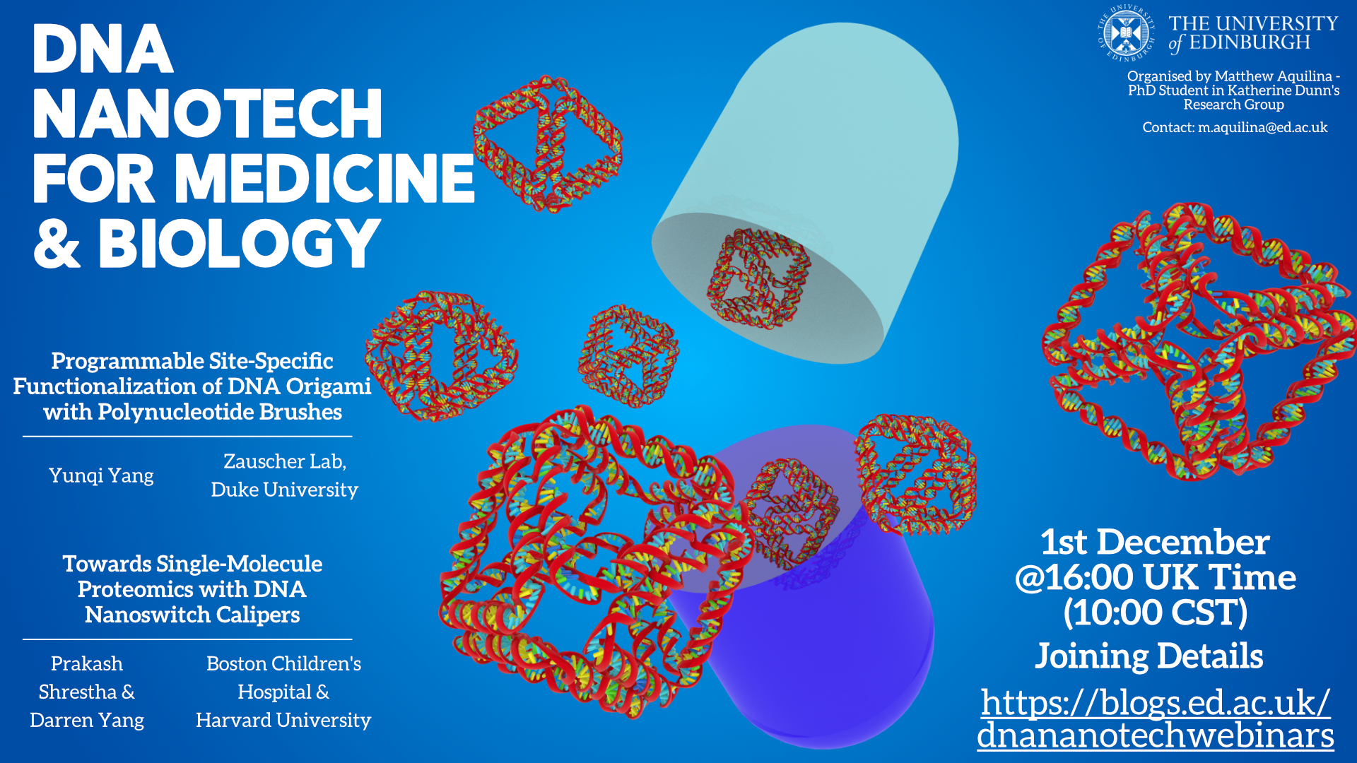
December 1st Watch Recording Here
- Yunqi Yang – Duke University
- Programmable Site-Specific Functionalization of DNA Origami with Polynucleotide Brushes
- Prakash Shrestha & Darren Yang – Boston Children’s Hospital & Harvard University
- Towards Single-Molecule Proteomics with DNA Nanoswitch Calipers
Talk Abstracts
Programmable Site-Specific Functionalization of DNA Origami with Polynucleotide Brushes: Our research is motivated by the need for stable, nanoparticle-based drug delivery platforms that are composed of nucleotides or nucleotide analogues. Towards this goal, we designed and synthesized site-specifically functionalized DNA origami nanostructures (DONs) using surface-initiated TdT-catalyzed enzymatic polymerization (SI-TcEP). This synergistic combination of surface-initiated enzymatic polynucleotide brush synthesis with precisely engineered DNA origami presents an innovative pathway for the generation of stable, polynucleotide brush functionalized origami nanostructures. Our experimental results show that we have precise control over multiple parameters, including the shape of the origami core, location, height, and composition of the brush. Coarse-grained oxDNA simulation provides us with a powerful tool to design and predict the morphology of origami-brush nanostructures. In addition, we have shown that polynucleotide-functionalized DONs have significantly higher nuclease resistance compared to unprotected DNA origami. Furthermore, this stability can be spatially programmed by designing the positions of the decorating sites on the surface of DONs, potentially allowing the control of the degradation of drug delivery vehicles to facilitate cellular uptake. Together, these attributes will enable new applications of DNA nanotechnology, such as in vivo molecular sensors and smart nanoscale delivery systems.
Towards Single-Molecule Proteomics with DNA Nanoswitch Calipers: Single-molecule distance measurements can provide an unprecedented view into the structure and dynamics of biomolecules and their interactions. However, current limitations in resolution and accuracy, and challenges in determining the spatial arrangement of multiple residues within each molecule, have restricted applications. By combining structural DNA nanotechnology with single-molecule force spectroscopy, we have developed a mechanically reconfigurable DNA Nanoswitch Caliper capable of measuring multiple coordinates on single biomolecules from heterogeneous samples with atomic resolution. Using dual-trap optical tweezers, we demonstrate absolute distance measurements with angstrom-level precision for both DNA and peptides, and using multiplexed magnetic tweezers, we demonstrate quantification of relative abundance in mixed samples. Measuring distances between DNA-labeled residues, we perform single-molecule fingerprinting of synthetic and natural peptides, and show discrimination, within a heterogeneous population, between different post-translational modifications. DNA Nanoswitch Calipers are a powerful and accessible tool for characterizing distances within nanoscale complexes that will enable new applications in fields such as single-molecule proteomics.
October 27th Watch Recording Here
- Nicolas Levy – ENS Lyon
- ENSnano: a Software for Designing 3D DNA Nanostructures
- Jurek Kozyra – CEO of Nanovery
- From Concept to Company: how to Build a DNA Nanotechnology Startup
Talk Abstracts
ENSnano: a Software for Designing 3D DNA Nanostructures: Since the 1990s, increasingly complex nanostructures have been reliably obtained out of self-assembled DNA strands: from “simple” 2D shapes to 3D gears and articulated nano-objects, and even computing structures. The success of the assembly of these structures relies on a fine tuning of their structure to match the peculiar geometry of DNA helices. Various softwares have been developed to help the designer. These softwares provide essentially four kind of tools: an abstract representation of DNA helices (e.g. cadnano, scadnano, DNApen, 3DNA, Hex-tiles); a 3D view of the design (e.g., vHelix, Adenita, oxDNAviewer); fully automated design (e.g., BScOR, Daedalus, Perdix, Talos, Athena), generally dedicated to a specific kind of design, such as wireframe origami; and coarse grain or thermodynamical physics simulations (e.g., oxDNA, MrDNA, SNUPI, Nupack, ViennaRNA,…). ENSnano is a new software for designing 3D DNA nanostructures that aims to conciliate all these approaches. It combines tried-and-tested features from other softwares and extend them with new exclusive functionalities. ENSnano offers a 3D live editable view synchronized with a 2D cadnano-like interface. By extending the concept of grids introduced in cadnano, ENSnano allows to abstract and articulate different parts of a 3D design. In this talk, we will make a state of the art of the currently available tools for designing DNA nanostructures. During this overview we will identify the key features of each software that made its success. We will then see how ENSnano gathers these functionalities in one centralized solution, and present ENSnano’s new features. Finally, we will discuss the main directions in which we want to extend ENSnano’s capabilities in the future and present our latest progress towards these goals.

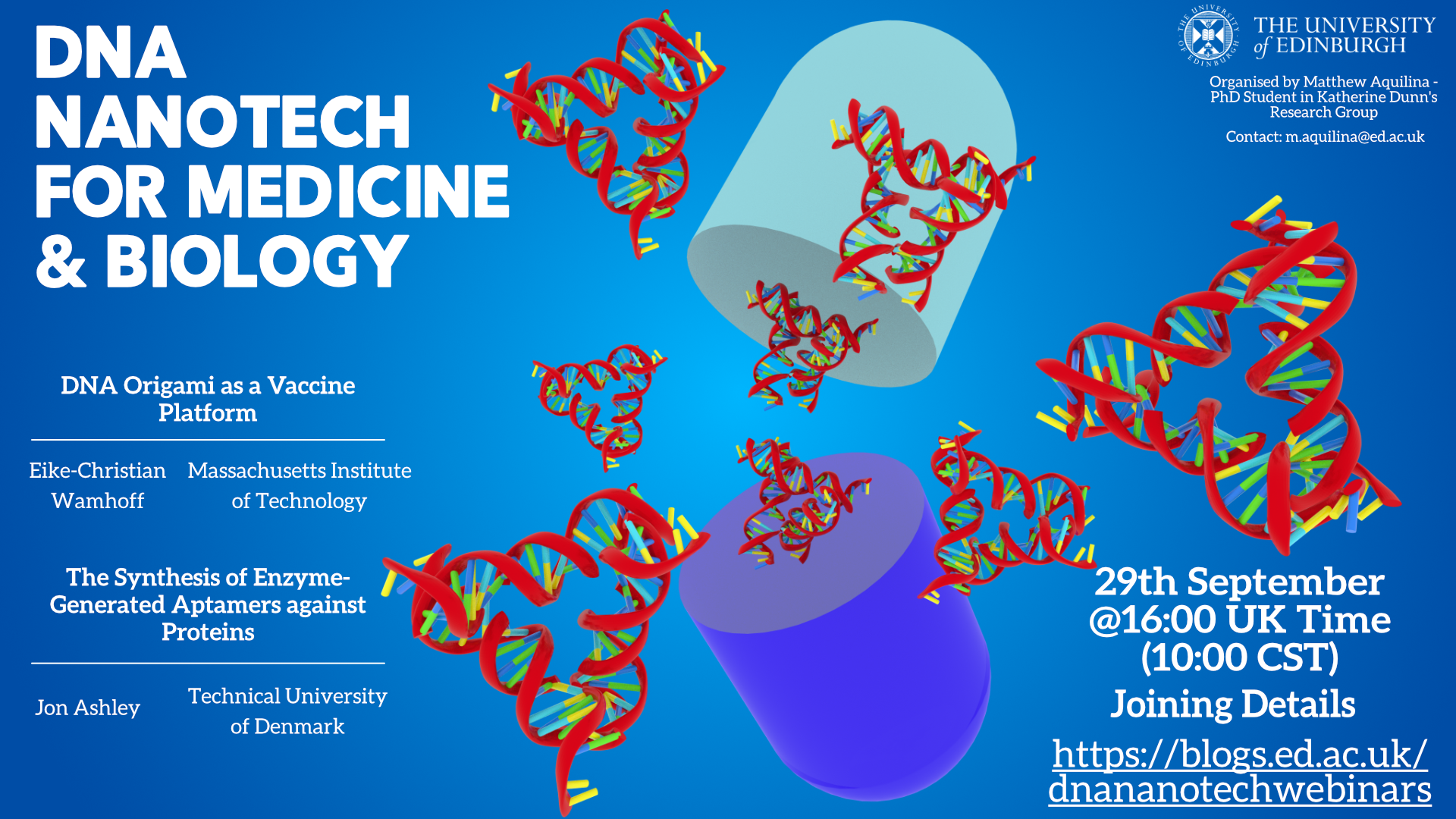
September 29th Watch Recording Here
- Eike-Christian Wamhoff – Massachusetts Institute of Technology
- DNA Origami as a Vaccine Platform
- Jon Ashley – Technical University of Denmark
- The Synthesis of Enzyme-Generated Aptamers against Proteins
Talk Abstracts
DNA Origami as a Vaccine Platform: Strategies to enhance antigen immunogenicity are of major importance to the design of subunit vaccines against infectious diseases, and may be critical for the generation of neutralizing antibodies (NAbs) for SARS-CoV-2 or HIV. One such strategy is the formulation of multivalent nanoparticles to promote B cell receptor (BCR) signaling and early B-cell activation. Here, DNA origami is particularly well-suited to precise organize antigens at the nanoscale while simultaneously enabling the screening of rationally designed, large-scale nanoparticles libraries due to its ease of formulation and modularity. We have recently defined the nanoscale parameters that drive BCR signaling in vitro. Here, I am presenting our progress towards validating this parameter set in vivo – based on advances in the formulation of DNA origami-based, multivalent subunit vaccines.
The Synthesis of Enzyme-Generated Aptamers against Proteins: Terminal deoxynucleotidyl transferase (TdT) is a unique polymerase enzyme capable of incorporating single nucleotides into an elongating polymer chain. The race to utilize this enzyme in polynucleotide synthesis in order to replace solid-phase DNA synthesis has grown over the last few years due to the low costs and possibility to synthesize polynucleotides beyond the current size limit beyond chemical solid phase techniques. Inspired by the methods used to generate molecularly imprinted polymers, we demonstrated for the first time, a new non-evolutionary approach for the synthesis of enzyme-generated aptamers capable of binding proteins with high affinity and selectivity. Enzyme-generated aptamers were synthesized by preparing a series of pre-polymerisation mixtures containing different ratios of nucleotides, oligonucleotide initiator sequences and incubating these mixtures with TdT. Through a feedback loop of controlling a number of parameters (ratios of nucleotides, initiator concentration and buffer conditions in each pre-polymerisation mixture and analyzing their apparent binding on a native gel, we were able to rationally design aptamers with high apparent binding towards each protein target. Bound enzyme generated aptamers were isolated, converted to dsDNA and sequenced. We demonstrated that candidate enzyme generated aptamers displayed specific multivalent binding towards their protein targets and this method allows us to screen by size as well as sequence, remove the current size caps placed on random libraries of ssDNA used in SELEX and minimize the PCR bias effect. As an extension, we also demonstrated that when a template protein is included into the pre-polymerisation mixtures before incubation with TdT enzyme, the apparent binding increases compared to a non-templated control, suggesting that we can drive the synthesis of enzyme-generated aptamers through a self-assembly based mechanism similar to molecular imprinting.
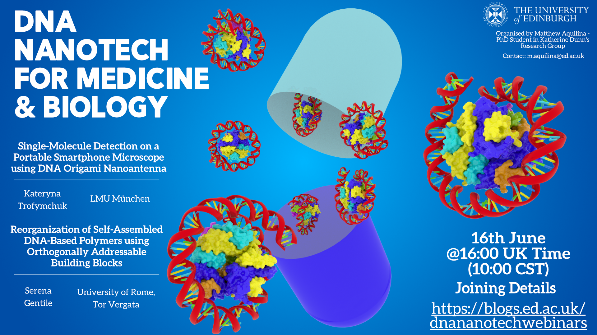
June 16th Watch Recording Here
- Kateryna Trofymchuk – LMU München
- Single-Molecule Detection on a Portable Smartphone Microscope using DNA Origami Nanoantenna
- Serena Gentile – University of Rome, Tor Vergata
- Reorganization of Self-Assembled DNA-Based Polymers using Orthogonally Addressable Building Blocks
Talk Abstracts
Single-Molecule Detection on a Portable Smartphone Microscope using DNA Origami Nanoantenna: Simplifying single-molecule detection would enable many exciting applications, such as point-of-care diagnostic. One strategy to achieve this ultimate sensitivity is to physically amplify fluorescence signals by plasmonic nanostructures. To address this question, we recently introduced NanoAntenna with Cleared HOtSpots (NACHOS). This is scaffolded by DNA origami nanostructure, that can be specifically tailored for the incorporation of bioassays and allows enhancing the fluorescence of a single-molecule up to 461-fold. This allowed us to carry out an exemplary single-molecule DNA detection on a portable, battery-powered smartphone microscope.
Reorganization of Self-Assembled DNA-Based Polymers using Orthogonally Addressable Building Blocks: Non-covalent interactions are the cornerstone upon which Nature has built the mechanisms to achieve dynamic reconfiguration of biopolymers and environmental adaptation. Emulating such properties and outcomes in rationally designed materials constitutes one of the main goals in the field of supramolecular chemistry. Successful approaches fulfilled the expectations using building-blocks capable of self-assembling in a predictable and versatile manner, which can also undergo through dynamic reconfigurations. Thanks to the highly programmable and specific non-covalent Watson-Creek interactions, synthetic DNA oligonucleotides have emerged as ideal candidates to configure supramolecular materials. Inspired by these observations we have explored a general strategy to control the reorganization of DNA-based polymers through external inputs. To do so, we have employed orthogonally addressable building blocks (i.e. DNA-tiles) that can self-assemble into tubular polymeric-like structures. These tiles share the same 5-nucleotide sticky ends responsible for self-assembly but differ in the sequence that can be orthogonally addressed by different regulator strands. The possibility to finely control (i.e. activate or inhibit) the tiles allowed to organize and reconfigure them into well-defined distributions: homopolymers, made of a single tile; random co-polymers, composed by different tiles distributed randomly and block structures, where the tiles are organized over specific portions of the polymers. The ease to engineer DNA-based addressable monomers opens new scenarios in the growing field of supramolecular polymers and paves the way to the possibility to reorganize them into multicomponent functional DNA structures.
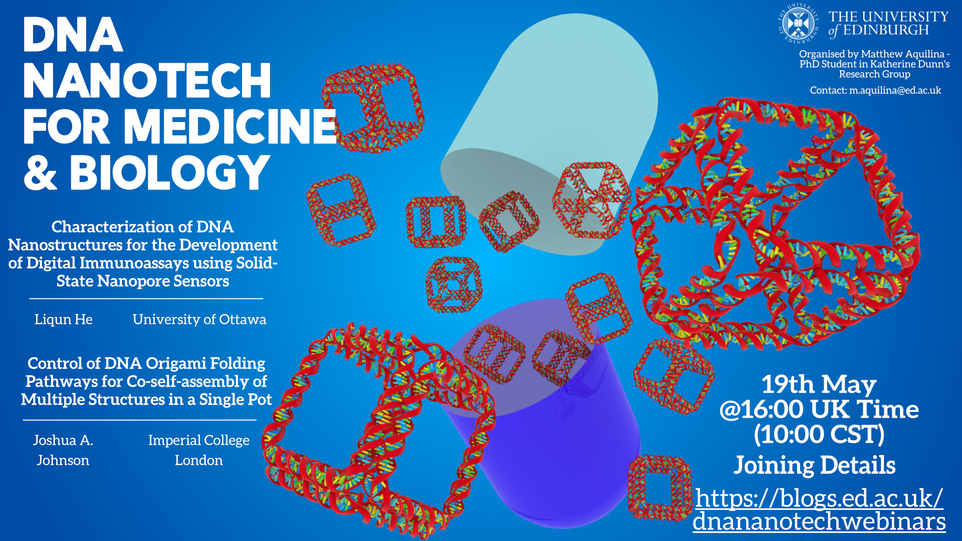
May 19th Watch Recording Here
- Liqun He – University of Ottawa
- Characterization of DNA Nanostructures for the Development of Digital Immunoassays using Solid-State Nanopore Sensors
- Joshua A. Johnson – Imperial College London
- Control of DNA Origami Folding Pathways for Co-self-assembly of Multiple Structures in a Single Pot
Talk Abstracts
Control of DNA Origami Folding Pathways for Co-self-assembly of Multiple Structures in a Single Pot: Self-assembly of DNA origami nanostructures has been shown to be primarily influenced by the entropic energy cost associated with the formation of scaffold loops and the kinetics of binding staple strands early in the self-assembly process. Here, we expand on these principles through investigations of co-self-assembly reactions, where we demonstrate the parameters necessary to fold two different nanostructures simultaneously in a single pot. We observe correlations between the kinetics of folding structures individually and the relative yields of structures in co-self-assembly reactions. These results demonstrate DNA origami folding proceeds along pathways that can be controllably bifurcated primarily by modulating parameters that alter folding rates. Furthermore, this work establishes a means of expanding the complexity of single-pot assemblies that contain multiple components for easier fabrication of complex systems.
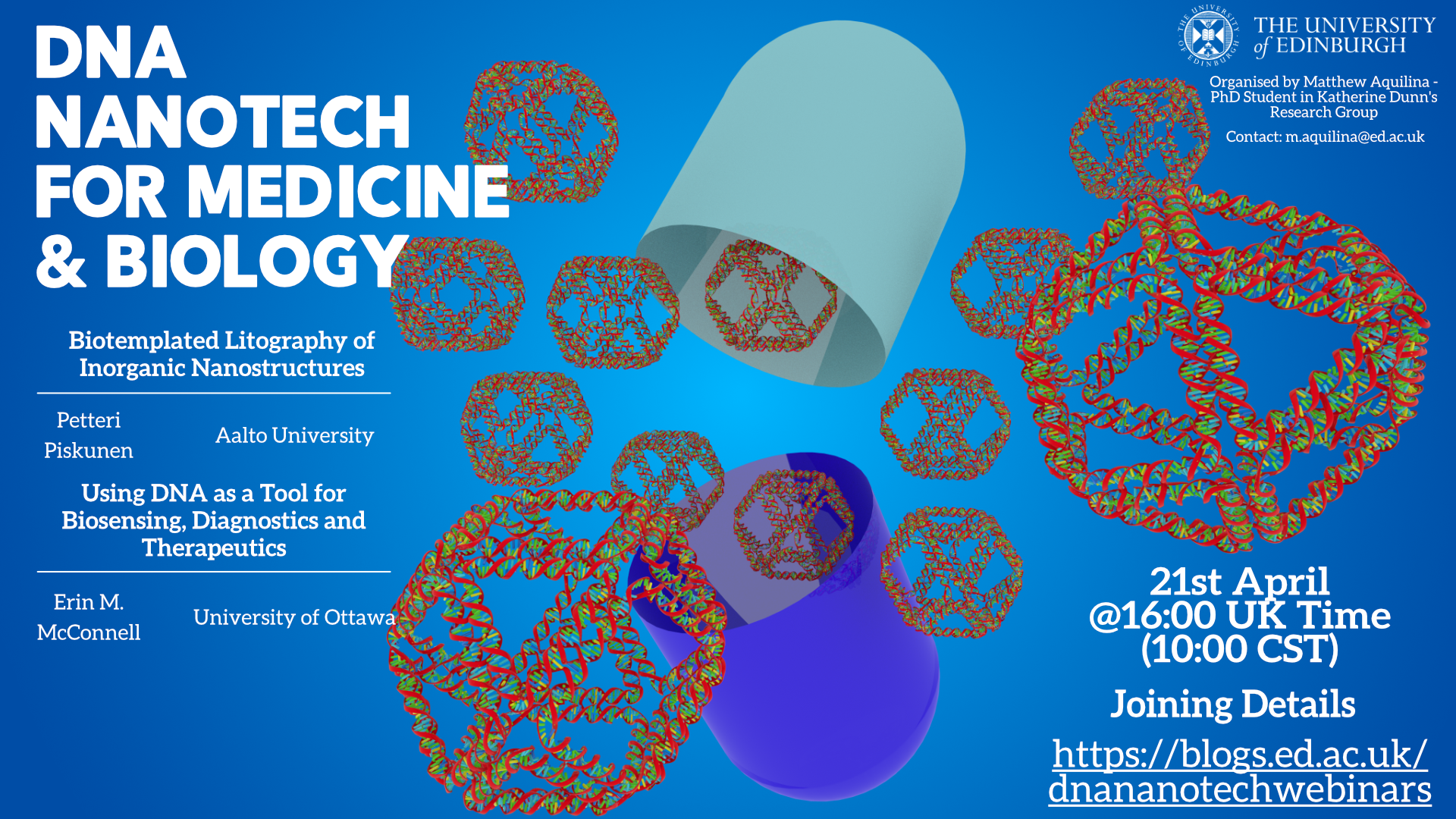
April 21st Watch Recording Here
- Petteri Piskunen – Aalto University
- Biotemplated Litography of Inorganic Nanostructures
- Erin M. McConnell – University of Ottawa
- Using DNA as a Tool for Biosensing, Diagnostics and Therapeutics
Talk Abstracts
Biotemplated Litography of Inorganic Nanostructures: One of the key benefits of DNA-based nanostructures is the amount of control that can be exerted over their shape. For example, with the DNA origami technique it is possible to use computer-aided design software to draft fully user-defined nanometer-precise 2D and 3D shapes, which can then be synthesized in a test tube at large scales. The resolution of these kinds of free form soft structures is far beyond the current limits of commercial solid-state nanofabrication techniques. Therefore, solid-state fabrication could progress beyond the state- of-the-art if DNA nanotechnology and prospectively other self-assembling biotemplates could be harnessed. One promising technique for this is the recently developed DNA-assisted lithography (DALI). The approach combines standard solid-state nanofabrication methods with DNA origami to transfer the shape of the origami into similarly shaped metallic nanostructures. While DALI can reach sub-10 nm feature sizes for the fabricated structures, it is only compatible with specialty substrates such as sapphire and silicon nitride.
Expanding on this concept, we present here a more generalized technique called biotemplated lithography of inorganic nanostructures (BLIN). The new technique circumvents the key material limitations of DALI, and furthermore, also enables the use of other biotemplates than DNA for lithographic patterning. We demonstrate the new technique by fabricating metallic (Ti, Au, Ag) and semiconducting (Ge) nanostructures on silicon and conductive glass substrates. The nanostructures are templated by DNA origami shapes with different geometries. We then demonstrate the use of a high-aspect-ratio tobacco mosaic virus (TMV) for creating micrometer-long Ag nanorods on silicon. The various geometries and aspect ratios highlight the potential and limitations of the current BLIN approach. With further optimization, the method could prove especially useful for the cost-effective manufacturing of optically active substrates for various plasmonic and biosensing applications.
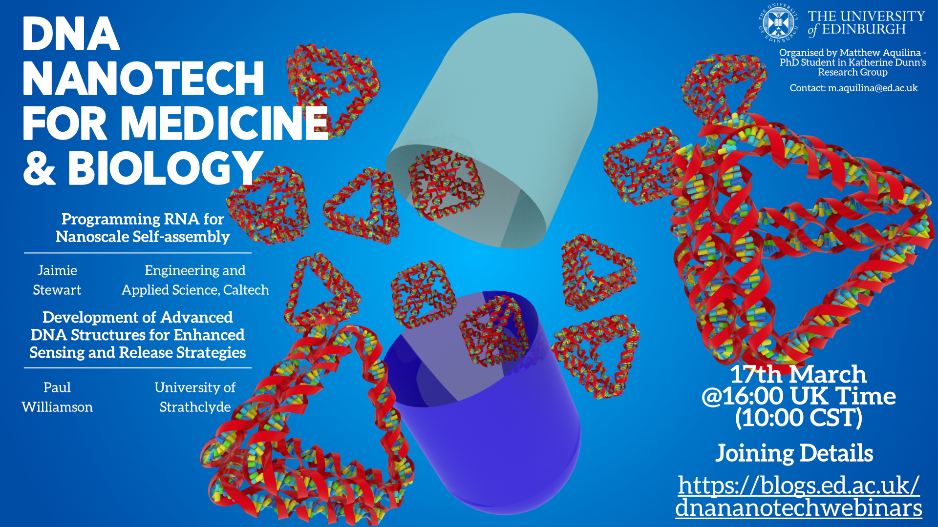
March 17th
- Jaimie Stewart – Engineering and Applied Science, Caltech
- Programming RNA for Nanoscale Self-assembly
- Paul Williamson – University of Strathclyde
- Development of Advanced DNA Structures for Enhanced Sensing and Release Strategies
Talk Abstracts
Programming RNA for Nanoscale Self-assembly: RNA is a functionally diverse molecule capable of interacting with molecules and responsible for regulating cellular activities. The canonical base pairing of RNA can be programmed for the self-assembly of architectures with nanoscale features of various size and complexity. Here, we demonstrate the self-assembly of multi-stranded RNA monomers into various architecture types. These assemblies are capable of both nanoscale organization and incorporation of functional RNA motifs to regulate gene expression or for the detection of other molecules.
Development of Advanced DNA Structures for Enhanced Sensing and Release Strategies: Electrochemical DNA biosensing systems are a critical emerging technology in the early detection of pathogens, and monitoring of human disease. In recent years there has been a growing interest in DNA sensing elements which go beyond linear probe strands, e.g. harpins and tetrahedra. This is because such structures can provide increased sensitivity, specificity and open up the possibility of combined sensing and release strategies. Here we present two ongoing studies, the first relates to the use of increasingly complex and redox tagged DNA probes to determine the presence of first target oligonucleotides, and second gene sequences for antimicrobial resistance. Electrochemical investigations by Differential Pulse Voltammetry (DPV) and Electrochemical Impedance Spectroscopy (EIS) of DNA Hairpin self-assembled monolayers demonstrated sensitivity to a sub nanomolar concentrations with a large dynamic range. The design of our hairpin provides enhanced specificity of target binding, with the structure capable of resolving single base pair mismatches in target sequences. Following on from this, we report a novel, and switchable, DNA origami interface. This study involved design, production and characterisation of a DNA origami ‘zipper’ structure, containing nine pH-responsive DNA locks. Each lock consisted of two parts, attached to the opposite arms of the zipper; a DNA hairpin and a single-stranded DNA that were able to form a DNA triplex through Hoogsteen base pairing. The sequences of the locks were selected so the zipper adopted a closed configuration at pH 6.5 and an open state at pH 8.0 (transition pKa 7.6). Thiolated versions of the zipper were immobilized onto gold electrodes and it was shown that the open and closed states could be probed electrochemically in a label free manner. These findings pave the way for development of DNA origami-based pH sensing and other pH-responsive sensing and release strategies for zipper-functionalised gold surfaces.
February 23rd
- Davide Michieletto – School of Physics and IGMM, University of Edinburgh
- Viscoelasticity of Topologically Active DNA
- Josie Kishi – Wyss Institute, Harvard University
- Using SABER to amplify multiplexed FISH and IF signals in situ
Talk Abstracts
Viscoelasticity of Topologically Active DNA: Polymer physics principles are increasingly acknowledged and applied to understand the behaviour of DNA organisation in vivo. Typically, they heavily rely on the assumption that the DNA does not change topology (or architecture) in time; in fact, this is hardly the case, as DNA is constantly topologically and mechanically re-arranged within the cell nucleus. Inspired by this, here I propose to study entangled fluids of DNA which can selectively alter their topology and architecture in time and may expend energy to do so. I argue that solutions of topologically active DNA in vitro can display unconventional viscoelastic behaviours and can be conveniently realised using solutions of DNA functionalised by certain families of proteins. In this talk I will present my first excursion into this field and present some recent results on the microrheology of entangled DNA undergoing digestion by restriction enzymes. I will present theories, simulations and experiments using particle tracking microrheology showing that that we can harness this non-equilibrium process to yield time-varying viscoelastic behaviours that may find application in controlled drug delivery.
Using SABER to amplify multiplexed FISH and IF signals in situ: Fluorescent in situ hybridization (FISH) and immunofluorescence (IF) techniques can provide quantitative information about the localizations and quantities of molecular species in fixed cells and tissues. However, challenges such as high tissue autofluorescence and slow imaging times due to long exposures continue to hinder our ability to map large tissues efficiently. Moreover, visualizing more than a few targets simultaneously can be tedious or impossible with standard methods. In this talk, I will introduce our newly developed signal amplification by exchange reaction (SABER) technique, which can be used to increase multiplexed signal levels from nucleic acid (SABER-FISH) or protein (Immuno-SABER) targets. Long, single-stranded DNA concatemers that are generated in vitro are applied simultaneously to targets of interest in situ to act as binding scaffolds for a multitude of short complementary strands conjugated to fluorophores (`imagers’). In different scenarios, we show that SABER can be used to amplify signal levels up to 450-fold, deployed against at least 17 targets simultaneously, and combined with expansion microscopy to achieve super-resolution imaging. The modularity, scalability, and cost-effectiveness of the SABER method make it a promising technique for future tissue mapping applications.